| The strange circle
| |||||||||
|
| |||||||||
|
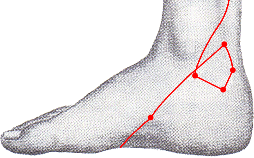
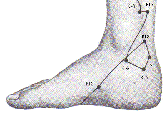
For years we interrogated skilled practitioners and scholars of Traditional Chinese Medicine (TCM) to know why the acupuncture meridian system (AMS) presents a circle in the kidney
meridian, precisely in the inner aspect of the ankle (picture above on
the left), and not elsewhere. When there was an answer, it was always the same: "Because books depict it like that". Lateral and oblique deviation, inverted direction, bends and crossings are common alterations of the meridians' path, but the circle in the ankle is the only circular line, out of the many metres the acupuncture system consists of. This is why
we defined it as strange. After comparing the acupuncture meridian charts with western anatomical and embryological knowledge, according to the findings reported in these pages,
we have formulated some working hypotheses:
| |||||||||
|
|
|||||||||
|
The observation |
|||||||||
|
|
| ||||||||
|
|
| ||||||||
|
|
| ||||||||
|
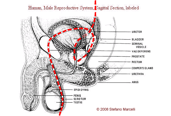
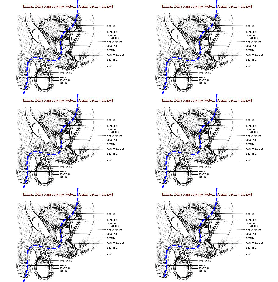
The urinary path is almost identical both in males and females, being different only in regard to the urethral length. |
| ||||||||
|
|
|||||||||
|
Comparative
anatomy |
|||||||||
|
|
| ||||||||
|
| |||||||||
|
|
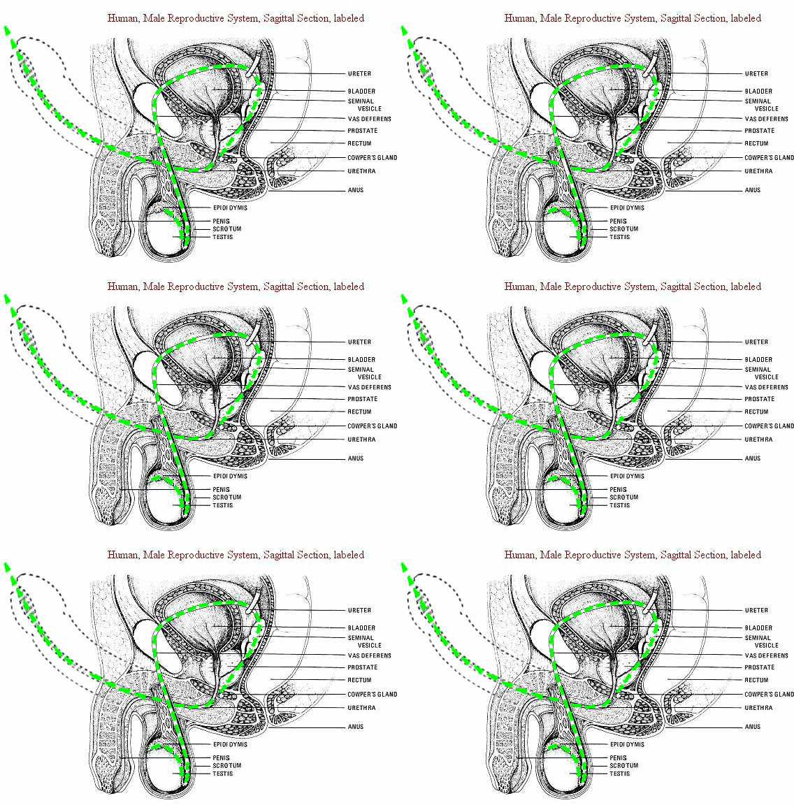
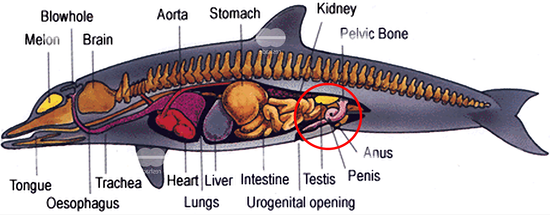
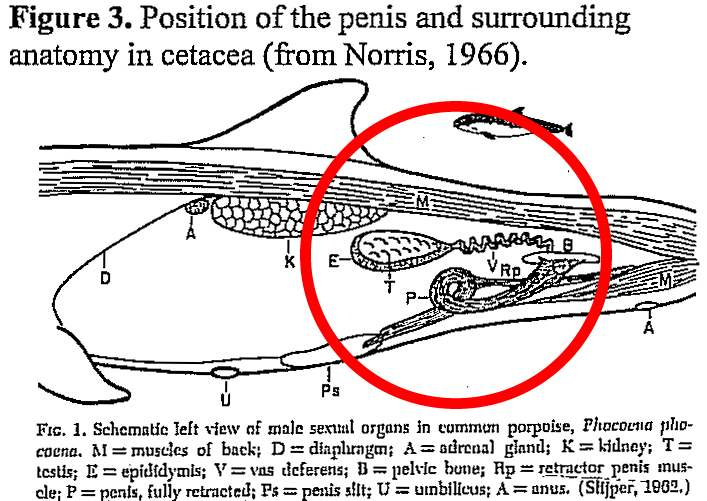
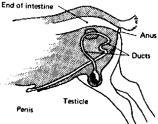
"The body of fibroelastic penis of the dolphin
[Norris, 1966] and sheep [May 1964] forms an s-shaped curve in the
retracted state (Fig. 3 and below). Erection involves straightening of
the s-curve and protrusion of the penis through a slit in the abdomen.
As there is no thickening or lengthening of the penis during erection,
rigidity depends more on the properties of the tunica albuginea
and trabeculae of the corpora cavernosa than on the
blood-filled cavernous spaces [Norris, 1966]. |
||||||||
|
|
|||||||||
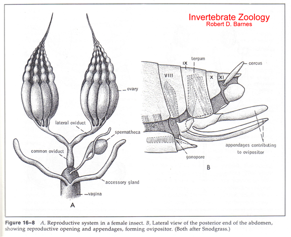 |
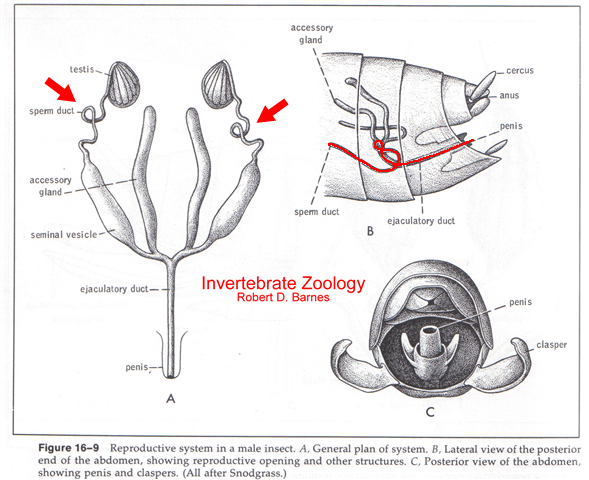
Insect Reproductive System The female reproductive system is shown in the drawing above on the left. The male is below. Variation among insect reproductive systems is great. Closely related species are often isolated from one another via small variations in the morphology of reproductive organs that prohibit interspecies mating. However, a generalized system can be constructed that closely represents all sexually reproducing insects. Be familiar with differences in male and female genitalia and be able to identify structures when given a diagram. Text taken from "Insect Morphology": University of Minnesota USA, Department of Entomology. Picture taken form "Invertebrate Zoology" by Robert B. Barnes. |
||||||||
 |
|||||||||
|
|
|||||||||
|
Embryology
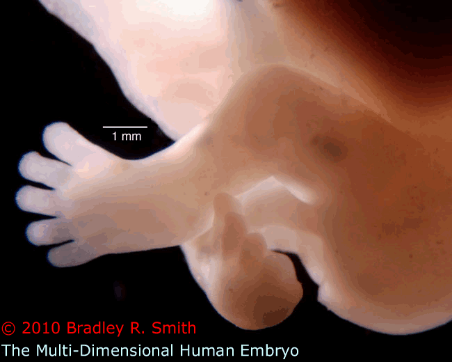
In the human embryo the proximity of the circle in the kidney meridian to the genital organs is remarkable. See also the picture at the title's right side (The Multi-Dimensional Human Embryo). The inner aspect of the ankle, where the circle is drawn, faces exactly the site where some time later testes (and not ovaries) will emerge.
Our principal question is: do the buds of the lower extremities also contain the strange circle in the kidney meridian? | |||||||||
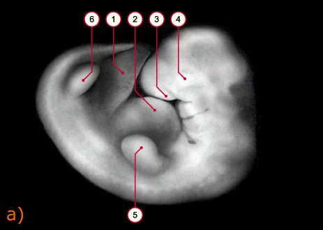
| a) © Professor Kohei Shiota, Kyoto University Human embryo at approx. 33th day, 14 mm. Characteristic signs: Lens vesicle covered with the ectoderm. Auditory primordium. Genesis of the hand plate. 1 Umbilical cord 2 Cardiac prominence 3 Nasal placode 4 Ocular primordium 5 Bud of the upper extremity 6 Bud of the lower extremity
|
 | ||||||||
|
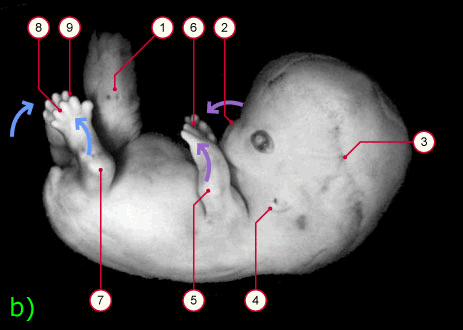
b) © Professor Kohei Shiota, Kyoto University Human embryo at approx. 51st day, 22-24 mm. Characteristic signs: Subcutaneous vessel network of the head spreads out. Hands and feet come closer and touch each other. 1 Umbilical cord with physiologic hernia 2 Nose 3 Subcutaneous vessel network of the head 4 Ear 5 Elbow 6 Pronation of the hands (pink arrow) 7 Knee 8 Supination of the feet (blue arrow) 9 Toes |
 | ||||||||
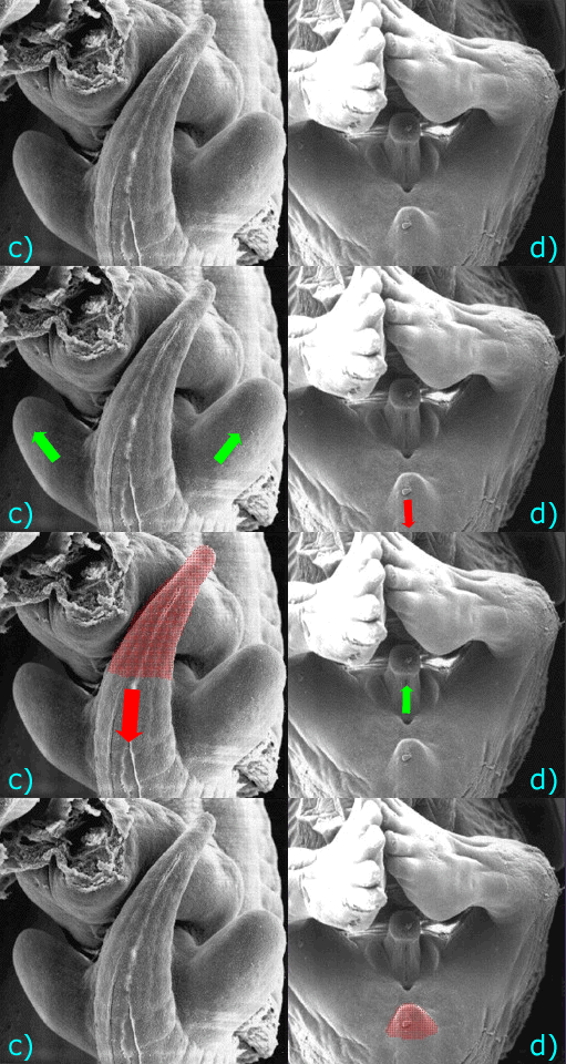
| c) and d) © MouseWorks, Inc. The forming external genitalia are not visible in this view of an 8 week human embryo on the left due to the prominent tail. The human tail regresses, as is evident in the 10 week embryo on the right. At the same time the lower limbs grow, the feet form, the external genital organs appears, and in males the testes travel along the inguinal channel toward the scrotum, their final destination. |
 | ||||||||
|
|
|||||||||
|
Scientific literature on
this specific subject There are a few works on the relation between acupuncture and embryology that corroborates with and are corroborated by our findings. ■
In 1989 Shang C. wrote the first
article that hypothesizes a relationship between acupuncture meridians and
embryonic morphogenesis. He proposed the involvement of the meridian system
in the growth regulation. "Both organizing centers and acupuncture points
have low electric resistance. The low electric resistance is related to the
distribution of gap junction and thus intercellular communication. Some
acupuncture points may be organizing centers. The meridian system is
important in coordination and regulation of morphogenesis. The properties of
organizing centers and acupuncture points can be explained in view of
singular point. Coupling and oscillation may underlie the mechanism of
acupuncture as well as growth regulation."
[1]. | |||||||||
|
|
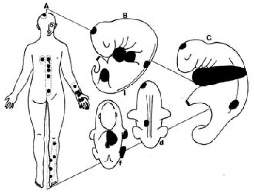
Acupuncture points with activity to cardiovascular pathology according to classic books, in adult (A), in a 6 weeks embryo (B): lateral (l) dorsal (d) and frontal view (f), and in a 4 weeks lateral view embryo (C). For a better comprehension in the embryological phases the cloud of localized points of the adult is represented as a black compact whole. | ||||||||
|
■
A very interesting work, corroborated by our findings and following theory, is the review of "Bonghan Channel System" by KS. Soh, physicist and researcher at Biomedical Physics Laboratory, Department of Physics and Astronomy, Seoul National University, Seoul, Korea
[9]. An excellent summary overview was made by David
Milbradt in 2009 [10]. | |||||||||
|
| |||||||||
|
References
[1] Shang C. Singular Point,
organizing center and acupuncture point. Am J Chin Med.
1989;17(3-4):119-27. | |||||||||
|
| |||||||||
|
Despite our western medical knowledge, since our first studies of TCM we have strongly felt that Acupuncture
Meridians must be real and not
abstract entities. But, year by years we became more and more skeptic
because of the lack of clear evidence, as far as we have begun to compare them
to gross anatomy. Our project's aim is to succeed in seeing the
acupuncture meridians in a scientific, technological and repeatable way.
If someone think this idea interesting and want to give his opinion is
invited to contact me.
Dr Stefano Marcelli, MD
| |||||||||
